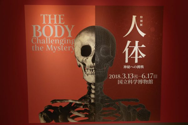
At the National Science Museum, a special exhibition "Human body – Challenge to mystery -" is held from March 13 (Tuesday) to Sunday, June 17, 2018 (Sun). Since the exhibition was held on March 12, I will report on that situation.
Our body is full of mystery. Why can we live and move? Human beings have challenged this mystery since the Renaissance era. In this exhibition, we look back on the achievements of pioneers such as Leonardo da Vinci and comment on the structure and function of the human body. In addition, the facts revealed by the latest technology will be introduced.
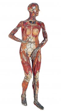
The human body model of Tamori who also served as MC for NHK Special "Huge network of human body mystery" is also …
How did mankind challenge "human body"?
· Leonardo da Vinci who dissected for painting
Leonardo da Vinci (1452 – 1519) who made a name as a painter performed human dissection with consent at a hospital in order to better depict the appearance of the extreme warrior appearing in "Battle of Anghiari" . When I started dissecting, he said that he considered the functions of the joints and the movement of the heart more than anatomists and grasped the structure of the human body in their own way.
Leonardo combines the findings obtained in dissection with the knowledge gained from the literature of his predecessors, and it is described in many pieces of paper. They were summarized in progeny and named "dissection manuscript".
Structure of the brain that Leonardo da Vinci thought

Cross section of the head from Leonardo da Vinci "dissection manuscript", part of the connection between the brain and eyes 1490 – 1992 Windsor Castle Royal Collection Collection
Royal Collection Trust / © Her Majesty Queen Elizabeth II 2018
Three round ventricles are drawn in the middle of the inside of the head. At that time, the ventricle was considered to be the center of brain functions filled with life vigor. This figure also shows how the optic nerve runs there. On the left side, an onion is drawn as a metaphor of the layer structure of the head.
· Human body model that contributed to dissemination of anatomy
■ Human body model with wax, wax model
Wax (wax) that has been considered a material of sculpture since ancient times. The wax model using it anatomically began to be created by Bologna craftsman, Gaetano Giulio Zumbo (1656-1701) at the end of the 17th century. However, since there was no way to preserve corpses in the 17th century, in order to continuously observe, it was necessary to autopsy dead bodies that always had freshness. About 200 body bodies were used to create a single wax model.
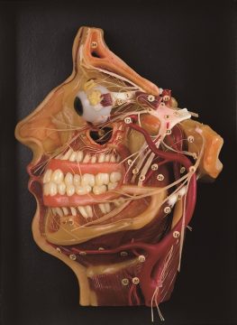
Head and neck wax model 19th century Japanese dental college medical doctor's museum collection
■ Human body model born as a teaching material, kinstrake
In the 19th century, an emphasis was placed on anatomical education, and the demand for human body model for education increased. Until then, the model was a wax model, but it was expensive and brittle making it unsuitable as a teaching material. Therefore, French anatomist Louis Tomah Jarome Ozu (1797-1880) devised a man model made of Pacheco. It was rich in durability and was able to disassemble and assemble, so it became very popular. It is imported to Japan via the Netherlands in the late Edo period.
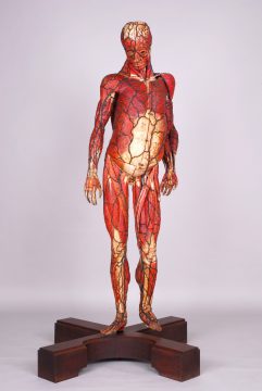
"Kinstraki" (Male) Collection of the Memorial Museum of the Kanazawa University School of Medicine, 19th Century
"Kinsutorureki" (female) Collection of the 19th century Fukui municipal museum of local history (From the 13th (Tue) to 17th May (Thursday) limited time display only)
Things that make people more crowded, brains
· Understanding the evolving brain
■ Network Theory … Is the nerve of the brain connected?
Italian doctor Camillo Golgi (1843-1926) developed a Golgi staining method using silver nitrate in 1873, succeeded in observing neurons of the brain. From this observation, Golgi asserted the "net-like theory" that the nerves of the brain are continuously connected and exhibit a reticular structure.
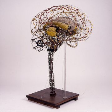
"Brain nerve fiber model" Switzerland, Buzi 1893 – 1910 Collection of the Boulhave Museum © Rijksmuseum Boerhaave,
Leiden V 25313
■ Neuron theory … The brain's nerve is not connected and some information transmission is done
Spanish doctor and neuroanatomist Santiago Ramón i · Cajar (1852-1934) used optical microscope Detailed observation asserted the neuron theory that "the nerves of the brain are discontinuously arranged and some information transmission is performed between adjacent cells".
In 1906, although both Golgi and Kahar awarded the Nobel prize at the same time, the lecture content at that seat was conflicting. After the death of the two names, the synapse clearance was confirmed using the electron microscope, so the army was raised in the neuron theory.
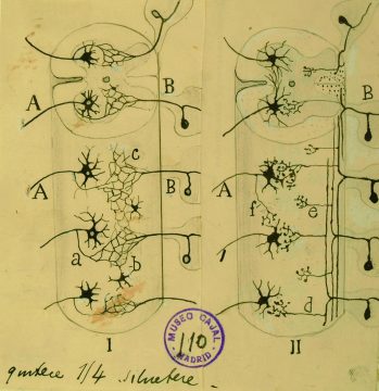
Contrast between net-like theory and neuron theory Santiago Ramón y · Kahar 1923 Collection of Kahal Research Institute
Cajal Institute, "Cajal Legacy", Spanish National Research Council (CSIC), Spain.
Schematic drawing drawn by Kaharu to explain the difference between Golgi's net-like theory (left in the figure) and neuron theory (right)
Human body research in the 21st century
· A new human body view overturning the concept of "brain's command tower"
With the advances in technology, the phenomena occurring in the body are now understood at the level of molecules and atoms. In the latest research, it is becoming clear that the preconceived notion that the brain is the commander of the whole body is being overturned, and it is becoming clear that all organs are helping each other while exchanging information directly without going through the brain.
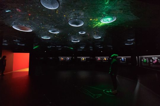
In this exhibition, 4K Super Hi-Vision images of the body are introduced, as well as a space expressing the inside of the body where the "messages" of the organs are fluttering.
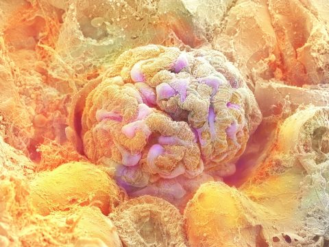
Kidney glomerulus © Daisuke Koka · Asahikawa Medical University / Hitachi High-Technologies / NHK
※ The picture is taken in rats. I put a color on the black and white image with an image.
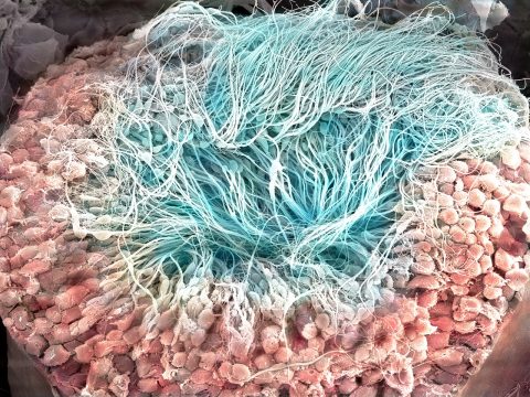
Testicular fine tube © Daisuke Koga · Asahikawa Medical University / NHK
※ The picture is taken in rats. I put a color on the black and white image with an image.
■ Network Symphony "Tired, tough" "The rice came!" … …. The conversation of the organs by "message substance" is in flight in our body. In "Network Symphony" of this exhibition, you can experience the noisy world inside the body with colors and sounds.
"Human body" which is the most familiar and at the same time a grand theme. Why do not you visit this exhibition and think about the mystery of our body.
Summary
| Exhibition name | Special exhibition "Human body – Challenge to mystery" |
| Venue | National Science Museum |
| A session | March 13 (Tue) – June 17 (Sun) in 2018 |
| closing day | Monday ※ Monday 26th (Monday), Monday April 2 (Monday), April 30 (Monday · Transfer holiday), June 11th (Monday) Open |
| time | 9 am – 5 pm (Friday and Saturday to 8 p.m.) ※ Admission until 30 minutes before closing time |
| Night Extension | 【Until 8:00 pm】 April 29 (Sun), 30 (month · transfer holiday), May 3 (Thu · holiday) 【until 6:00 pm】 May 1 (Tue), 2 (Wednesday), 6 (Sun) |
| Price | General · College students: 1,600 yen Small, middle and high school students: 600 yen |
| URL | http://jintai2018.jp |


![]()